

3 December 2024
Understanding Key Radiologic Imaging Techniques: Which Methods Are Best for Different Conditions?
Explore key radiologic imaging techniques and learn which methods are most effective for diagnosing different medical conditions.
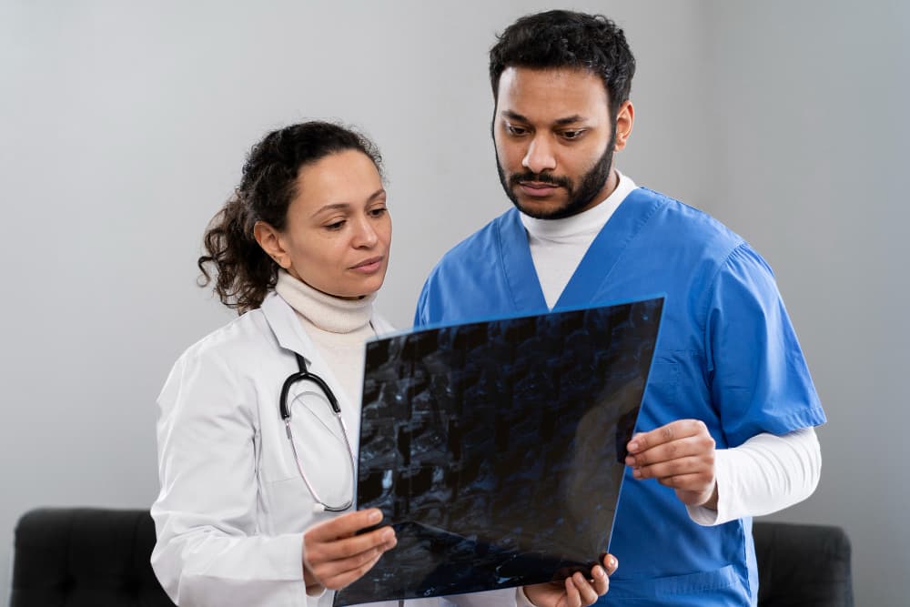
Radiologic imaging has transformed the field of medical diagnostics, offering a range of imaging techniques that allow healthcare providers to visualise the internal structures of the body without invasive surgery. Each imaging modality has unique applications, advantages, and limitations. This article dives into the most commonly used radiologic imaging techniques, examining the specific benefits and uses of each method.
What Are the Most Common Radiologic Imaging Techniques?
Radiologic imaging encompasses several primary techniques, each suited to visualising different body structures and conditions. Here’s a list of commonly used imaging techniques, their applications, and what makes each unique:
- X-ray Imaging: X-rays are quick, cost-effective, and commonly used to examine bone fractures and lung conditions. They use low doses of ionising radiation to create images.
- Computed Tomography (CT) Scan: CT scans use a combination of X-rays and computer technology to create detailed cross-sectional images, offering a more comprehensive view than a standard X-ray.
- Magnetic Resonance Imaging (MRI): MRIs produce high-resolution images of soft tissues, such as muscles and the brain, without using radiation, relying instead on magnetic fields.
- Ultrasound Imaging: This technique uses sound waves rather than radiation, making it particularly safe for applications such as prenatal monitoring and soft tissue imaging.
- Positron Emission Tomography (PET) Scan: PET scans provide functional imaging by using radioactive tracers, often used in oncology to detect cancerous cells.
- Fluoroscopy: Offering real-time imaging, fluoroscopy is ideal for guiding certain procedures and examining moving structures, like the gastrointestinal tract.
- Nuclear Medicine Imaging: By using radioactive isotopes, nuclear medicine visualises the function and structure of organs, frequently used for thyroid and cardiac assessments.
What Are the Key Attributes of Radiologic Imaging Techniques?
Each radiologic imaging technique carries distinct attributes that make it appropriate for specific diagnostic needs. When selecting an imaging technique, healthcare providers consider factors such as radiation exposure, resolution, procedure time, and the body area to be examined.
| Imaging Technique | Primary Use | Radiation Exposure | Pros | Cons |
| X-ray | Bone fractures, chest | Low | Quick, cost-effective | Limited soft tissue visibility |
| CT Scan | Internal injuries, tumours | Moderate | Detailed cross-sectional views | Higher radiation, costly |
| MRI | Soft tissue, brain, spine | None | High-resolution soft tissue | Expensive, lengthy procedure |
| Ultrasound | Pregnancy, organs | None | Safe, no radiation | Limited detail for dense organs |
| PET Scan | Cancer, metabolic activity | Moderate | Functional information | Requires radioactive tracers |
| Fluoroscopy | Real-time procedures | Moderate | Continuous imaging, real-time | Higher radiation, specialised use |
| Nuclear Medicine | Organ function, bones | Moderate to high | Functional assessment | Involves radioactive isotopes |
Which Radiologic Imaging Techniques Are Best for Specific Conditions?
Selecting the correct imaging modality depends on the specific condition being examined. Here’s how different imaging techniques align with various health assessments:
- Bone Fractures: X-rays are the standard approach, offering fast and clear imaging for diagnosing fractures.
- Soft Tissue Injury or Tumour Detection: MRI provides superior soft tissue resolution, ideal for detecting tumours, brain abnormalities, and joint injuries.
- Internal Injuries and Complex Fractures: CT scans are optimal for complex injuries as they provide a multi-angle view of structures.
- Pregnancy and Soft Organ Imaging: Ultrasound, being radiation-free, is ideal for foetal imaging and for examining organs such as the liver or kidney.
- Oncologic Applications: PET scans are invaluable in identifying cancer cells and monitoring response to treatment, providing functional insights that anatomical imaging alone cannot achieve.
What Considerations Should Be Taken Into Account When Choosing an Imaging Technique?
Several factors influence the choice of imaging modality:
- Purpose of Exam: Whether it’s for diagnosis, monitoring, or guiding treatment influences the choice.
- Body Area or Organ to be Scanned: Each technique has strengths for specific anatomical areas.
- Need for Functional Data: Techniques like PET scans and nuclear medicine offer insight into organ function, unlike purely anatomical methods.
- Patient Condition: Factors like claustrophobia, mobility issues, or implanted devices can impact the choice.
- Preparation Requirements: Some techniques require fasting or avoiding metal, particularly for MRI and certain nuclear medicine procedures.
| Technique | Best For | Limitations | Preparation Needed | Time Required |
| X-ray | Bones, chest imaging | Limited soft tissue detail | None | Minutes |
| CT Scan | Trauma, soft tissue | Higher radiation than X-rays | Contrast dye for some | Minutes to an hour |
| MRI | Soft tissue, brain, spine | Cannot be used with metal implants | Avoid metal | 20-60 minutes |
| Ultrasound | Organs, foetal imaging | Limited tissue penetration | None | Minutes |
| PET Scan | Cancer, brain activity | Requires radiotracer injection | Fasting required | 30-60 minutes |
| Fluoroscopy | GI tract, catheter placement | Continuous radiation exposure | Contrast dye for some | Minutes |
| Nuclear Medicine | Organ function | Radiation exposure from isotopes | Fasting in some cases | Variable |
How Does Radiation Exposure Vary Between Techniques?
Radiation exposure is a significant factor when choosing an imaging technique. While X-rays and CT scans involve low to moderate radiation, MRI and ultrasound are radiation-free. PET scans and nuclear medicine procedures use radiotracers, providing valuable functional information but with radiation exposure, which is generally moderate.
For example:
- CT Scans provide higher radiation levels than standard X-rays due to the detailed cross-sectional imaging.
- MRI and Ultrasound are safe alternatives, particularly important for patients requiring multiple scans, such as those with chronic conditions.
- Nuclear Medicine procedures use radioactive isotopes; these are carefully managed to balance diagnostic needs and patient safety.
How Do Costs Differ Across Radiologic Imaging Techniques?
Costs vary widely due to differences in technology, procedure time, and availability:
- X-rays are generally inexpensive and widely available.
- CT Scans and MRIs are more expensive due to the complex machinery and specialised interpretation required.
- PET Scans and Nuclear Medicine procedures are high-cost because of radiotracer production and the need for specialised equipment.
Is Preparation Required for Radiologic Imaging?
Some imaging techniques have specific preparation protocols:
- CT Scans often require a contrast dye to enhance visibility.
- MRI involves removing all metal objects due to the magnetic field.
- PET Scans and Nuclear Medicine may involve fasting or avoiding certain medications.
- Ultrasound may require a full bladder for clearer pelvic images.
Preparation guidelines are provided to ensure optimal image quality and patient safety. Always follow pre-scan instructions carefully.
What Research Supports These Radiologic Techniques?
Current evidence supports the use of specific imaging techniques for accurate diagnosis and treatment planning. The Royal Australian and New Zealand College of Radiologists provides guidelines for using different imaging modalities based on condition and patient demographics. According to recent studies, MRI is especially useful in diagnosing brain and spinal conditions, while PET scans have proven effective in oncology (source: RANZCR).
Research has also underscored the importance of limiting radiation exposure, especially in children, for whom MRI and ultrasound are preferred (source: Australian Government Department of Health).
At Border Radiology Specialists, we’re committed to providing the highest standards in radiologic imaging. Our specialists are trained to help you choose the most appropriate imaging technique based on your medical needs, ensuring both safety and accuracy. We offer a comprehensive range of services tailored to meet the unique needs of each patient.
Reference Section
- Royal Australian and New Zealand College of Radiologists
- Australian Government Department of Health
- Radiation Protection of Patients – Australian Radiation Protection and Nuclear Safety Agency
FAQs
- What is the difference between MRI and CT scans?
MRI uses magnetic fields to create high-resolution images of soft tissues, while CT scans use X-rays to create cross-sectional images, making them ideal for visualising bone and certain internal injuries. - Are there any risks associated with radiologic imaging?
Most radiologic imaging techniques are safe when conducted appropriately. However, certain methods, such as CT scans and PET scans, involve radiation exposure, which should be limited when possible. - Can I eat or drink before an imaging procedure?
Preparation depends on the specific procedure. CT scans may require contrast, and PET scans often require fasting. Consult your radiologist for precise guidelines. - Which imaging technique is safest for children?
MRI and ultrasound are preferred for children, as these methods do not use radiation, unlike X-rays or CT scans. - What imaging technique is best for cancer detection?
PET scans are commonly used for cancer detection and monitoring, as they provide functional information about metabolic activity, which helps identify cancer cells. - How long does an MRI scan take?
An MRI can take anywhere from 20 to 60 minutes, depending on the area being examined. The process is painless, but patients must remain still for optimal image quality.
This article is general information, not a substitute for medical advice. For guidance about your own situation, speak with your GP or call us on (02) 6046 9755.