

3 December 2024
Understanding Central Skull Base Lesions: What Are the Key Imaging Insights for Better Diagnosis?
Discover key imaging insights for diagnosing central skull base lesions, and understand how advanced techniques enhance accuracy in radiologic assessments.
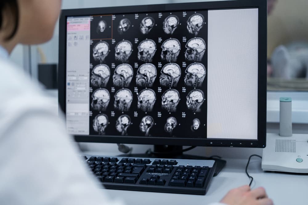
The central skull base is an anatomically complex region that can present a range of lesions, from benign growths to aggressive malignant tumours. Effective diagnosis and management of these lesions require advanced imaging techniques, particularly MRI and CT scans. In this article, we will delve into the different types of central skull base lesions, their imaging features, and how specific imaging techniques can help in identifying and differentiating these complex structures. With thorough knowledge of these insights, clinicians can provide more accurate diagnoses, better treatment planning, and improved outcomes for patients.
What Are Central Skull Base Lesions and Their Types?
Central skull base lesions are a diverse group of abnormalities that arise from or affect the bones and tissues at the skull base. This region contains essential structures, including cranial nerves and blood vessels, that connect the brain to the rest of the body. Due to the density of critical structures, central skull base lesions often cause neurological symptoms, and their management requires careful imaging assessment.
Types of Central Skull Base Lesions
Central skull base lesions can be broadly classified into the following categories:
- Tumours
- Meningiomas: Often benign, arising from the meninges.
- Chordomas: Rare, slow-growing, and originating from the notochord remnants.
- Chondrosarcomas: Malignant tumours originating from cartilage.
- Pituitary Adenomas: Tumours of the pituitary gland, with some having suprasellar extension.
- Inflammatory Lesions
- Sinusitis: Inflammation of the sinus, sometimes extending to skull base structures.
- Osteomyelitis: Infection of bone, potentially involving the skull base.
- Vascular Malformations
- Aneurysms: Abnormal bulging of blood vessels in the skull base region.
- Arteriovenous Malformations (AVMs): Tangled blood vessels bypassing normal brain tissue.
- Cysts and Developmental Lesions
- Rathke’s Cleft Cysts: Non-cancerous cysts near the pituitary gland.
- Epidermoid and Dermoid Cysts: Slow-growing cystic lesions arising from ectodermal remnants.
- Infectious Lesions
- Abscesses: Localised infections that can develop in or near the skull base.
- Fungal Infections: Infections in immunocompromised individuals that can extend to the skull base.
Each type of lesion displays unique imaging characteristics, which are essential for differential diagnosis and developing an effective treatment strategy.
What Imaging Modalities Are Essential for Skull Base Lesions?
Correct imaging modality selection is crucial for central skull base lesions. MRI and CT are the primary imaging techniques used due to their high-resolution capabilities and complementary roles. Other modalities, like PET and ultrasound, can offer additional information, particularly in assessing metabolic activity and soft-tissue characteristics. Here is a summary of the common imaging modalities used for skull base lesions:
| Imaging Technique | Strengths | Limitations |
| MRI | High soft tissue contrast, multiplanar views | Time-consuming, contraindications (e.g., pacemaker) |
| CT | Fast, good for bone detail, readily available | Limited soft tissue contrast, radiation exposure |
| PET-CT | Detects metabolic activity, useful in malignancy | Limited spatial resolution, high cost |
| Ultrasound | No radiation, good for superficial lesions | Limited in bony areas, not useful for deeper lesions |
| Angiography | Excellent for vascular lesions | Invasive, risk of complications |
MRI and CT: The Cornerstones of Skull Base Imaging
MRI is the preferred modality for soft tissue resolution, particularly in distinguishing different types of brain and soft-tissue lesions. CT imaging, on the other hand, provides detailed bone assessment, which is invaluable for evaluating bony erosion or calcification patterns commonly seen in chondrosarcomas and chordomas. PET-CT may also be used in cases where there’s a need to identify malignant activity or recurrence.
What Are the Key Imaging Features for Skull Base Lesions?
The unique imaging characteristics of skull base lesions, as seen on MRI or CT, provide essential clues that guide diagnosis. Common imaging features include lesion location, growth patterns, signal intensity on MRI or density on CT, and enhancement patterns with contrast. Below is a breakdown of these findings by lesion type:
| Lesion Type | Common Imaging Findings | Imaging Modality |
| Meningioma | Hyperintense on T2 MRI, Calcifications, Dural Tail | MRI, CT |
| Chordoma | Bone erosion, midline location, high signal on T2 MRI | MRI, CT |
| Chondrosarcoma | Calcifications, lobulated mass, adjacent bone erosion | MRI, CT |
| Pituitary Adenoma | Enlarged sella, contrast enhancement, suprasellar extension | MRI |
| AVM | Flow voids on MRI, tortuous vessels, high-flow signal | MRI, Angiography |
| Abscess | Ring enhancement, restricted diffusion | MRI |
| Epidermoid Cyst | Low density on CT, high T2 signal, no enhancement | MRI, CT |
By analysing these imaging features, radiologists can narrow down differential diagnoses. For instance, a lesion with a dural tail on MRI is often indicative of a meningioma, while high T2 signal in a midline lesion with bone erosion could suggest a chordoma. Accurate diagnosis often relies on the integration of imaging data with clinical findings and patient history.
How Does Differential Diagnosis Aid in Identifying Skull Base Lesions?
Differentiating between benign and malignant lesions in the skull base is essential for determining appropriate management. Certain imaging features can hint at whether a lesion is likely benign or malignant. For example, benign lesions generally have slow growth rates, well-defined borders, and minimal bone involvement, while malignant lesions exhibit aggressive growth, infiltrative borders, and extensive bone destruction.
| Feature | Benign Lesion Characteristics | Malignant Lesion Characteristics |
| Growth Rate | Slow | Fast |
| Borders | Well-defined, non-infiltrative | Irregular, infiltrative |
| Calcifications | Common (e.g., meningiomas) | Less common |
| Signal Intensity (MRI) | Homogeneous, less enhancement | Heterogeneous, marked enhancement |
| Bone Involvement | Usually minimal | Often invasive with bone destruction |
| Associated Symptoms | Mild or absent | Progressive, neurological deficits |
Through differential diagnosis, radiologists can evaluate these imaging findings to rule out benign versus malignant conditions, leading to a more targeted and effective treatment plan.
How Are Central Skull Base Lesions Managed Once Diagnosed?
Management of central skull base lesions varies based on lesion type, location, and severity. Treatment options can range from surgical resection and radiotherapy to medical management in cases of infectious or inflammatory lesions. Treatment planning often depends on the comprehensive assessment of lesion type and size, as well as the patient’s overall health and symptoms. Advanced imaging is used in treatment planning to guide precision surgery, radiotherapy, or conservative management.
How Important Is Timely Diagnosis and Imaging?
Timely diagnosis through imaging is critical to managing central skull base lesions effectively. Prompt imaging can identify complications like bone erosion, cranial nerve compression, and intracranial extension, which can significantly impact patient outcomes. MRI, with its high tissue contrast, can reveal subtle abnormalities, while CT can detect early signs of bone involvement. For complex or recurrent lesions, repeat imaging may be needed for ongoing assessment, ensuring that the treatment approach remains appropriate and effective.
Border Radiology Specialists can provide expert imaging support for those dealing with central skull base lesions. With advanced imaging modalities and a team dedicated to precise, patient-centred care, our specialists are equipped to assist clinicians in making accurate diagnoses for better patient outcomes.
References
- The Royal Australian and New Zealand College of Radiologists. (n.d.). Understanding Skull Base Imaging: A Clinical Guide. Retrieved from https://www.ranzcr.com
- Australian Institute of Health and Welfare. (2023). Neuroimaging in Australia: A Statistical Review. Retrieved from https://www.aihw.gov.au
- The Royal Melbourne Hospital. (2022). Advanced Imaging Techniques in Skull Base Lesions. Retrieved from https://www.thermh.org.au
Frequently Asked Questions
- What are common symptoms of central skull base lesions?
Symptoms include headaches, vision changes, hearing impairment, cranial nerve palsies, and facial pain. - Which imaging modality is most effective for identifying skull base tumours?
MRI is typically the best for assessing soft tissue, while CT provides superior bone detail, making both essential for a full evaluation. - How do benign and malignant skull base lesions differ in imaging?
Benign lesions usually have well-defined borders and slow growth, while malignant lesions often appear invasive with rapid growth and irregular margins. - Can skull base lesions be treated non-surgically?
Yes, non-surgical options like radiotherapy and medical management are available, especially for inflammatory and infectious lesions. - What role does differential diagnosis play in skull base lesion assessment?
Differential diagnosis helps distinguish between lesion types, leading to more accurate diagnosis and targeted treatment. - Is repeat imaging necessary for skull base lesions?
In cases of progressive lesions or those under treatment, follow-up imaging may be needed to monitor changes and adapt the treatment plan.
This article is general information, not a substitute for medical advice. For guidance about your own situation, speak with your GP or call us on (02) 6046 9755.