

3 December 2024
The Role of MRI and CT in Diagnosing Pancreatic Cysts: A Radiologist’s Approach
Explore the role of MRI and CT in diagnosing pancreatic cysts, with insights from a radiologist’s approach to accurate imaging and diagnosis.
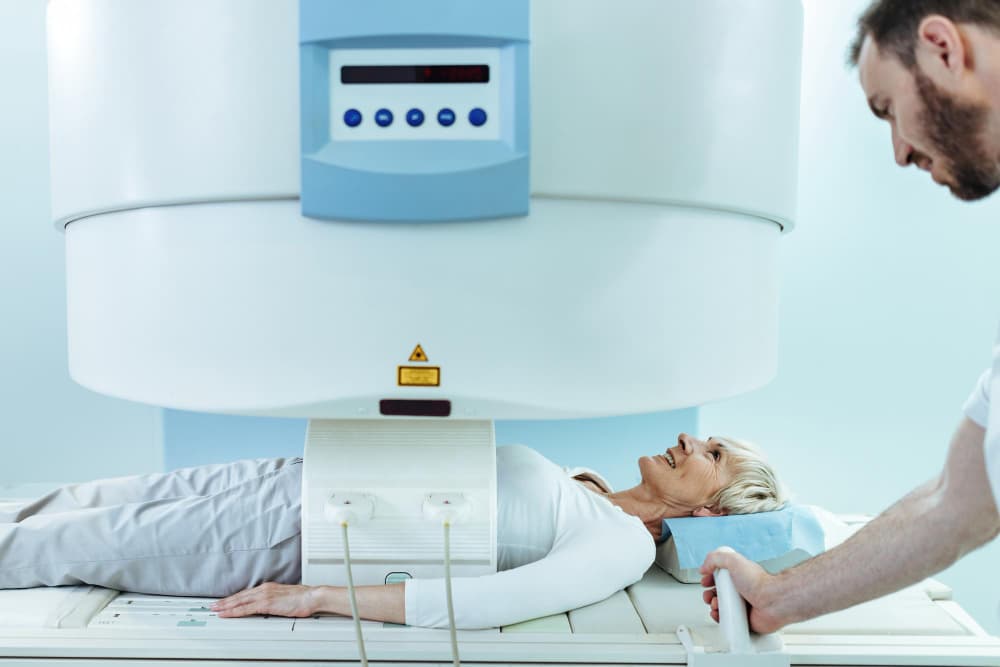
Pancreatic cysts are becoming increasingly common, largely due to advancements in imaging techniques that allow for clearer views of pancreatic lesions. Identifying these cysts accurately is crucial for patient outcomes, as some cyst types carry a risk of malignancy, while others are benign. Radiologists primarily rely on MRI and CT imaging to evaluate these cysts, each of which has unique strengths in diagnosis. In this article, we explore the role of MRI and CT in diagnosing pancreatic cysts, focusing on how each modality supports a radiologist’s approach.
What Makes MRI Essential for Pancreatic Cyst Evaluation?
MRI (Magnetic Resonance Imaging) is often preferred for its high soft-tissue contrast, allowing for better visualisation of fluid-filled and solid components within pancreatic cysts. Unlike CT, MRI does not involve ionising radiation, making it suitable for repeat imaging, particularly in younger or high-risk patients. MRI’s ability to characterise the internal structure of cysts, such as septations, nodules, and the cyst wall thickness, is critical for differential diagnosis.
The following attributes make MRI ideal for pancreatic cyst evaluation:
- High Soft-Tissue Contrast: MRI excels in distinguishing between different types of soft tissues. For example, it can highlight fluid levels in cysts on T2-weighted images and detect any solid components.
- Signal Characteristics: MRI uses T1, T2, and Diffusion-weighted imaging sequences, enhancing the details seen within a cyst, which is essential in differentiating benign from potentially malignant lesions.
- Gadolinium-Based Contrast: In cases where contrast is needed, gadolinium-enhanced MRI provides insights into the vascularity of a cyst. Enhancing solid areas within a cyst may suggest malignancy.
- No Radiation Exposure: For patients requiring long-term monitoring, MRI is preferred due to the absence of radiation.
Radiologist’s Checklist for MRI Pancreatic Cyst Assessment:
- Cyst size, location, and shape
- Morphology (unilocular, multilocular, septated)
- Cyst contents (solid, fluid-filled, calcified)
- Communication with pancreatic ducts
- Enhancement pattern following contrast administration
How Does CT Complement MRI in Pancreatic Imaging?
CT (Computed Tomography) is known for its high spatial resolution, allowing radiologists to see detailed anatomical structures and locate cysts within the pancreas more precisely. CT is also valuable in initial evaluations when the cyst’s nature is unknown, as it can quickly identify features such as calcifications and wall thickness. CT is often chosen in emergency settings due to its rapid acquisition time. However, it does involve ionising radiation, which limits its use in repeat examinations.
Key CT attributes include:
- High Spatial Resolution: CT imaging captures clear anatomical details, which is particularly helpful in locating cysts, understanding their relationship to nearby structures, and identifying calcifications.
- Contrast-Enhanced Phases: CT scans utilise iodine-based contrast agents that provide distinct arterial, venous, and delayed phase images, essential for detecting vascularised lesions.
- Quick Acquisition Time: CT scans are completed within seconds, making them advantageous in acute care settings.
Radiologist’s Checklist for CT Pancreatic Cyst Assessment:
- Cyst’s anatomical location and size
- Presence of calcifications (indicative of serous cystadenomas)
- Wall and septal thickness
- Degree of vascularity through iodine-based contrast
- Communication with main pancreatic duct (a common feature in IPMNs)
What Are the Imaging Characteristics of Different Pancreatic Cyst Types?
Each type of pancreatic cyst exhibits specific imaging characteristics that radiologists use to determine its nature. By knowing the typical features of each cyst type, a radiologist can tailor their diagnostic approach to maximise accuracy and recommend appropriate follow-up. Below is a comparative table detailing how MRI and CT capture different cyst features.
MRI vs. CT for Diagnosing Pancreatic Cysts
| Attribute | MRI | CT |
| Radiation Exposure | None | High |
| Contrast Agents | Gadolinium-based | Iodine-based |
| Image Resolution | High soft-tissue contrast | High anatomical detail |
| Ideal for | Soft-tissue and fluid composition | Structural details and calcifications |
| Limitations | Higher cost, longer acquisition | Radiation exposure, limited tissue contrast |
| Sensitivity to Cyst Components | Excellent for fluid and septa | Better for calcifications |
| Suitability for Surveillance | Excellent for follow-up | Suitable for initial diagnosis |
Common Types of Pancreatic Cysts and Imaging Features
| Cyst Type | MRI Features | CT Features |
| Serous Cystadenoma | Honeycomb appearance, low T1, high T2 signal | Thin-walled, possible calcifications |
| Mucinous Cystic Neoplasm | High T2 signal, septations, wall enhancement | Unilocular or multilocular, wall enhancement |
| Intraductal Papillary Mucinous Neoplasm (IPMN) | Ductal dilation, gadolinium enhancement | Ductal dilation, contrast-enhancing septa |
| Solid-Pseudopapillary Neoplasm | Mixed signal, solid and cystic areas | Partially solid, possible calcifications |
| Simple Cysts/Pseudocysts | Uniform T2 high signal, non-enhancing wall | Well-defined, thin-walled, homogeneous |
What Factors Influence the Choice of MRI or CT?
The choice of MRI or CT depends on various factors, including the patient’s health, the cyst’s characteristics, and the goal of imaging. Below are some considerations that guide the selection of modality:
- Patient’s Health: MRI is preferred for patients requiring ongoing surveillance due to its non-ionising nature. However, CT is favoured in acute settings where rapid diagnosis is crucial.
- Cyst Characteristics: MRI is better for differentiating cyst contents, while CT provides a detailed anatomical context, especially useful for complex cyst structures or those with calcifications.
- Cost and Accessibility: MRI is generally more costly and less accessible than CT, which can influence decisions in certain healthcare settings.
- Diagnosis vs. Surveillance: MRI is typically used for long-term monitoring, while CT may be more practical for initial diagnosis.
How Do MRI and CT Contribute to Differential Diagnosis of Pancreatic Cysts?
Differentiating between benign and malignant cysts is a primary concern for radiologists. Each cyst type has distinguishing features that may suggest a higher or lower risk of malignancy. Understanding these characteristics is essential in forming an accurate diagnosis.
Differential Diagnosis of Pancreatic Cysts Based on Imaging
| Imaging Characteristic | Possible Diagnosis | Modality with Highest Sensitivity |
| Unilocular cyst without enhancement | Simple Cyst/Pseudocyst | MRI |
| Multilocular cyst with septations | Mucinous Cystic Neoplasm (MCN) | MRI |
| Ductal dilation with cystic areas | Intraductal Papillary Mucinous Neoplasm (IPMN) | MRI |
| Cyst with internal calcifications | Serous Cystadenoma | CT |
| Solid and cystic composition | Solid-Pseudopapillary Neoplasm | CT and MRI |
Which Modality Should Be Used for Monitoring Pancreatic Cysts?
In cases where cysts are benign but need monitoring due to their potential for malignant transformation, MRI is the preferred modality. The absence of ionising radiation allows for periodic imaging without increasing the patient’s risk. MRI also allows radiologists to track changes in the cyst’s size, morphology, and internal composition over time.
What Research Supports the Use of MRI and CT for Pancreatic Cysts?
Numerous studies support the specific roles of MRI and CT in the imaging and differential diagnosis of pancreatic cysts. According to a 2022 study in Pancreatology, MRI was shown to be particularly effective in differentiating between serous and mucinous cysts, with a diagnostic accuracy exceeding 85%. Other studies in European Radiology highlighted the role of MRI in identifying high-risk IPMNs due to its sensitivity to ductal dilation and mural nodules. CT was validated as an effective initial screening tool due to its quick acquisition and high anatomical detail, with particular effectiveness in identifying calcified lesions.
For patients seeking detailed and careful imaging for pancreatic cysts, Border Radiology Specialists offers expert radiological services tailored to each patient’s diagnostic needs. Our team is dedicated to providing state-of-the-art MRI and CT imaging, ensuring accurate diagnoses and quality patient care.
References
- Pancreatology Journal (2022). Diagnostic Efficacy of MRI vs CT in Pancreatic Cyst Evaluation. Retrieved from Pancreatology.com.
- European Radiology Journal (2021). Advances in MRI Imaging for Intraductal Papillary Mucinous Neoplasms. Retrieved from EuropeanRadiology.com.
- Royal Australian and New Zealand College of Radiologists (2023). Best Practice Guidelines for Pancreatic Cyst Imaging. Retrieved from RANZCR.org.
FAQs
- What are the main differences between MRI and CT for pancreatic cyst diagnosis?
MRI offers superior soft-tissue contrast and is ideal for differentiating cystic contents. CT, however, provides detailed anatomical resolution, especially useful for detecting calcifications.
- Is MRI always better than CT for pancreatic cyst monitoring?
MRI is preferred for long-term monitoring due to the absence of radiation, but CT can be useful for initial diagnostic assessments.
- What are the radiation risks associated with CT imaging for pancreatic cysts?
CT involves ionising radiation, which can be a concern, especially for patients requiring repeated scans. MRI, in contrast, uses no radiation.
- Are there any risks associated with gadolinium-based contrast in MRI?
Gadolinium is generally safe but may pose risks for patients with kidney problems. Radiologists assess individual risks before administering contrast.
- How often should benign pancreatic cysts be monitored?
Surveillance frequency depends on the cyst type and size, often ranging from 6 to 12 months. A radiologist will recommend a schedule based on individual risk factors.
- Can both MRI and CT be used together for a more accurate diagnosis?
Yes, combining MRI and CT can provide a comprehensive view, especially in complex cases or when cyst characteristics are ambiguous.
This article is general information, not a substitute for medical advice. For guidance about your own situation, speak with your GP or call us on (02) 6046 9755.