

3 December 2024
Reducing Radiation Exposure: What Are the Best Tips for Safer Imaging?
Explore effective tips for reducing radiation exposure in medical imaging, ensuring safer practices while maintaining accurate results.
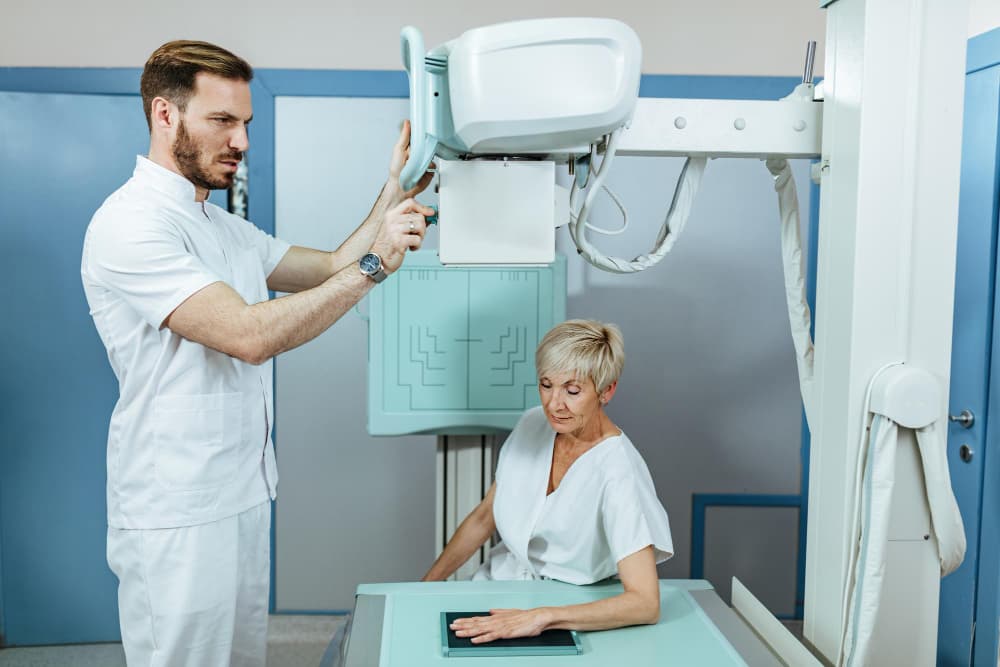
Medical imaging, encompassing X-rays, CT scans, mammograms, and fluoroscopy, has revolutionised diagnostics, enabling healthcare professionals to identify and address conditions with precision. However, many of these imaging methods use ionising radiation, which, in high or repeated doses, can pose risks. This article explores how radiation exposure in imaging can be minimised for optimal patient safety, following principles like ALARA (As Low As Reasonably Achievable) and offering practical strategies for both practitioners and patients.
What Are the Most Effective Tips to Reduce Radiation Exposure?
Reducing radiation exposure in medical imaging is critical, especially when alternative, lower-dose or non-ionising options may be available. Here are essential safety tips:
- Use the ALARA Principle: Minimising exposure is always the goal, achieved by reducing the dose or frequency of scans, especially for high-dose imaging like CT scans.
- Consider Non-Ionising Alternatives: Where possible, MRI or ultrasound imaging, which do not involve ionising radiation, should be the preferred choice.
- Utilise Shielding Equipment: Lead aprons, thyroid shields, and other protective devices can significantly reduce radiation exposure to sensitive areas.
- Regularly Calibrate Imaging Equipment: Routine calibration ensures that imaging devices provide accurate, low-dose scans without compromising quality.
- Tailor Imaging Settings to Patient Needs: Adjust the dose based on factors like age, body size, and medical history, particularly for vulnerable groups.
- Educate Patients and Staff: By informing patients about potential risks and the purpose of each imaging procedure, you reduce anxiety and enhance cooperation for necessary safety measures.
What Are Safe Imaging Techniques and Technologies?
Different imaging methods offer varying levels of radiation exposure. While X-rays and CT scans are common and effective, there are safer imaging techniques that can limit patient exposure without compromising diagnostic quality. Below is a comparison of radiation exposure levels across imaging techniques and some safe practices to consider.
| Imaging Technique | Radiation Level (mSv) | Common Uses | Notes on Safety |
| X-Ray | 0.1 – 1.5 | Bone fractures, chest exams | Use shielding; optimise settings for specific regions. |
| Computed Tomography (CT) | 1 – 20 | Full body scans, detailed organ imaging | Low-dose options available; use for essential cases. |
| Mammography | 0.4 | Breast cancer screening | Low exposure; follow screening guidelines. |
| Fluoroscopy | 1 – 10 | Gastrointestinal exams, catheter placements | Utilise pulsed mode; limit to essential cases. |
| MRI | 0 (non-ionising) | Soft tissue, neurological exams | Ideal for frequent scans and soft tissue views. |
| Ultrasound | 0 (non-ionising) | Pregnancy, soft tissue, blood flow | Safe for repeated exams; suitable for many applications. |
What Are Non-Ionising Imaging Alternatives?
Non-ionising imaging techniques, such as MRI and ultrasound, do not expose patients to radiation, making them suitable for sensitive groups, including children and pregnant women. These methods are especially effective for soft tissue imaging, providing detailed images without the risks associated with ionising radiation.
Non-ionising techniques offer a safe alternative for follow-up examinations, reducing cumulative exposure risks over time. Magnetic Resonance Imaging (MRI), for instance, is frequently used for neurological assessments, while ultrasound is ideal for monitoring pregnancies and evaluating soft tissues or blood flow.
How Does Patient Education Help Reduce Radiation Exposure?
Patient education plays an essential role in radiation safety. By understanding the necessity of each scan and the precautions being taken, patients are often more receptive to safety measures like shielding and adjusting positioning. Here are some key points to communicate to patients:
- Explain the ALARA Principle: Patients benefit from understanding why minimising radiation exposure is a priority, reducing concerns and fostering compliance with safety practices.
- Highlight the Benefits of Shielding: Inform patients of the value of shielding measures, such as lead aprons, for safeguarding vulnerable areas.
- Encourage Record-Keeping of Imaging History: Patients who keep a record of past imaging procedures can share this with healthcare providers to avoid unnecessary repeat scans.
- Promote Awareness of Non-Ionising Options: Inform patients when MRI or ultrasound may be suitable alternatives to ionising imaging methods, especially for routine or follow-up scans.
Why Are Professional Standards Important in Imaging Safety?
Professional standards guide radiology practices, helping practitioners achieve safe imaging protocols. Key principles include:
- Adherence to ALARA: This foundational principle emphasises minimising radiation by reducing dosage and frequency wherever possible.
- Diagnostic Reference Levels (DRLs): These reference values provide benchmarks for typical radiation doses in medical imaging, helping ensure that doses remain within safe limits.
- Regular Compliance Checks and Training: Routine staff training and equipment calibration support consistent application of radiation safety measures.
What Are the Key Safety Practices for Different Patient Demographics?
Different patient groups require unique safety measures due to varying sensitivities and risk factors. Children, pregnant women, and elderly patients are particularly vulnerable to radiation, necessitating tailored approaches.
| Patient Demographic | Safety Measures | Notes on Exposure Reduction |
| Children | Use paediatric protocols, limit scan areas | Children are highly sensitive; minimise exposure rigorously. |
| Pregnant Women | Opt for ultrasound or MRI where possible | Avoid ionising radiation unless essential. |
| Elderly Patients | Reduce scan frequency and dose | Monitor cumulative exposure, especially in chronic cases. |
| General Population | ALARA practices, shield use, low-dose settings | Optimise dosage and educate on necessity of each imaging. |
How Can Imaging Techniques Reduce Cumulative Radiation Exposure?
The goal of imaging is to obtain the clearest possible diagnostic information with minimal risk. Cumulative radiation exposure, however, is a growing concern, particularly with frequent scans or high-dose imaging. Techniques to reduce cumulative exposure include:
- Dose Modulation: Techniques like iterative reconstruction and adaptive statistical reconstruction allow lower doses without compromising image quality.
- Pulsed Fluoroscopy: Unlike continuous fluoroscopy, pulsed mode reduces exposure by emitting radiation in intervals, effective in procedures like catheter placements.
- Low-Dose CT Protocols: Specialised low-dose protocols can produce effective results for specific exams, such as lung screenings.
- Digital Radiography: Digital X-rays allow lower radiation doses compared to conventional film-based radiography, helping reduce exposure over time.
Why Is Routine Calibration of Imaging Equipment Essential?
Regular calibration ensures that imaging equipment produces accurate results at the lowest possible dose. Calibration maintains equipment efficiency, reduces the risk of excessive radiation exposure due to malfunction, and supports adherence to regulatory standards. Equipment that is not routinely calibrated may emit inconsistent doses, affecting both patient safety and image quality.
How Does Shielding Protect Patients During Imaging?
Protective shielding is an important component of radiation safety, especially for vulnerable areas like the thyroid and reproductive organs. Lead aprons, thyroid shields, and lead glasses are commonly used in X-rays and CT scans. Shielding reduces radiation exposure to non-targeted parts of the body, allowing the focused area to receive the necessary dose without risking nearby organs.
Australian regulatory bodies strongly advocate for the use of shielding, especially in procedures involving high-dose imaging, to ensure patient safety. Studies indicate that the use of lead aprons in chest X-rays reduces exposure to organs outside the primary imaging area by over 90% (source: Australian Radiation Protection and Nuclear Safety Agency).
For professional imaging services where patient safety is a priority, visit Border Radiology Specialists. We provide comprehensive and safe imaging options, utilising advanced technology and following stringent safety protocols to ensure optimal care for every patient.
Reference Section
- Australian Radiation Protection and Nuclear Safety Agency (ARPANSA). “Guidelines for Minimising Radiation Exposure.” Available at: ARPANSA
- Royal Australian and New Zealand College of Radiologists. “Standards of Practice for Diagnostic and Interventional Radiology.” Available at: RANZCR
FAQs
What is the ALARA principle?
The ALARA principle stands for “As Low As Reasonably Achievable,” and it’s a guideline in radiology for minimising radiation exposure by optimising imaging settings, reducing scan frequency, and utilising protective measures like shielding.
Can non-ionising imaging alternatives replace all X-rays and CT scans?
While non-ionising methods like MRI and ultrasound are safer for certain tissues and follow-up exams, they may not always provide the same level of detail for specific conditions, particularly in bone imaging or complex internal assessments.
How can I keep track of my radiation exposure history?
You can maintain a personal record of all imaging procedures, including the type of scan and date. Sharing this with healthcare providers helps avoid unnecessary repeat scans, reducing cumulative radiation exposure.
What is a low-dose CT scan, and is it as effective as a regular CT?
Low-dose CT scans use lower radiation levels, especially beneficial for routine screenings like lung checks. While they are often effective, they may not provide the same clarity in detailed assessments, making them suitable for specific conditions but not all cases.
Are there age restrictions on certain imaging techniques?
No strict age restrictions exist, but children are more sensitive to radiation. Paediatric-specific protocols or non-ionising imaging are recommended for this demographic to minimise exposure risk.
What should I ask my doctor about radiation exposure before an imaging procedure?
You can ask if the procedure is essential, whether there are lower-dose options or non-ionising alternatives, and if protective shielding will be used.
This article is general information, not a substitute for medical advice. For guidance about your own situation, speak with your GP or call us on (02) 6046 9755.