

3 December 2024
Recognising Normal Skeletal Variants in Radiology: What Do They Mean and How Are They Identified?
Learn to recognize normal skeletal variants in radiology, understand their significance, and how they are identified in imaging.
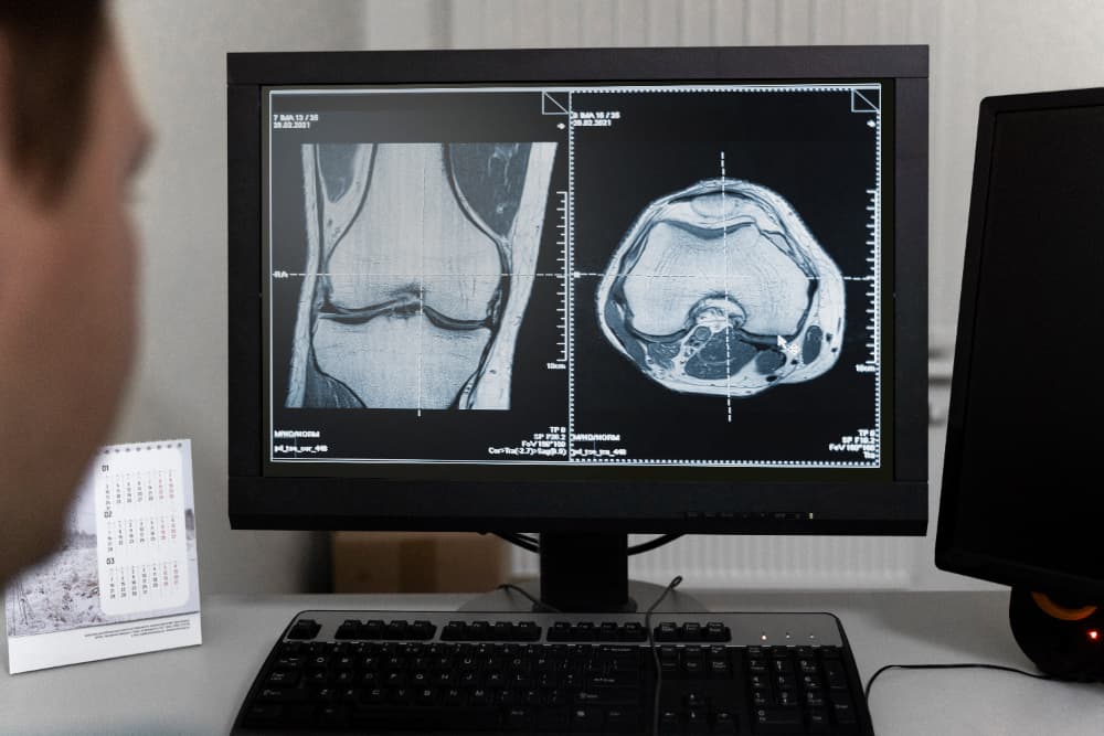
Radiologists frequently encounter skeletal anomalies, or “variants,” that appear on imaging. These are common deviations from textbook anatomy that don’t typically indicate disease or injury but can complicate the reading of radiographic images. Recognising normal skeletal variants is critical to ensure that radiologists and clinicians accurately distinguish between a benign anomaly and a pathological finding.
This article explores common skeletal variants, their characteristics, and diagnostic imaging techniques, as well as how radiologists can identify them confidently to avoid misdiagnosis.
What Are the Most Common Normal Skeletal Variants?
Skeletal variants include a range of anatomical deviations that can affect bones, joints, and cartilage. They vary significantly in presentation and location, often appearing in the feet, spine, and other areas. Below is a list of some of the most frequently encountered skeletal variants in radiology:
- Accessory Ossicles – These are small, extra bone pieces commonly found near joints, such as the os trigonum in the foot.
- Vertebral Anomalies – Includes conditions like sacralisation of L5 or lumbarisation of S1, where the vertebrae have altered structures.
- Bifid Ribs – A forked rib structure often observed as an incidental finding.
- Sesamoid Bones – Small bones within tendons, such as the fabella behind the knee.
- Supracondylar Process of the Humerus – A bony projection on the humerus, which can sometimes cause impingement of nerves.
- Bipartite Patella – A kneecap divided into two parts.
- Calcaneal Variants – Variations of the calcaneus bone, often showing as an extra facet.
- Cervical Rib – An additional rib arising from the seventh cervical vertebra, which can sometimes cause thoracic outlet syndrome (TOS).
How Are Skeletal Variants Diagnosed in Radiology?
Recognising skeletal variants often involves understanding which imaging modalities best display these anomalies and distinguishing them from pathological findings. Here are the primary imaging modalities used:
- X-ray: This remains the primary tool for observing bone density, shape, and structural anomalies.
- CT Scan: Used for high-detail imaging, especially useful in complex bone structures or when a variant is suspected.
- MRI: Effective for detecting soft tissue differentiation around skeletal variants.
- Ultrasound: Occasionally used for imaging around joints or tendons impacted by variants, particularly useful for sesamoid bones.
Each modality provides different levels of detail, helping radiologists make accurate assessments and avoid over-diagnosing benign anatomical variants as disease.
What Are Some Normal Skeletal Variants and Their Characteristics?
Below is a table of common skeletal variants, their anatomical locations, radiographic appearances, and clinical significance. Recognising these helps radiologists avoid misinterpretation and ensures that benign findings are not confused with potential pathologies.
| Variant | Location | Radiographic Appearance | Frequency | Clinical Significance |
| Accessory Ossicles | Foot, hand | Small, round bone fragments | Common in population | Usually incidental, rare symptoms |
| Cervical Rib | C7 vertebra | Bony projection from C7 | Rare | May cause thoracic outlet syndrome |
| Bifid Rib | Rib cage | Forked rib appearance | Rare | Generally asymptomatic |
| Bipartite Patella | Patella (knee) | Two-part patella | 1-2% of population | Incidental, may cause discomfort |
| Supracondylar Process | Humerus | Small bony spur | Rare | May impinge on median nerve |
| Sesamoid Bones | Joints (e.g., knee, thumb) | Small round bones within tendons | Variable | Often asymptomatic |
How Do You Differentiate Skeletal Variants from Pathological Findings?
Differentiating between normal variants and pathological findings is essential to ensure accurate radiographic analysis. This differentiation is especially important for certain conditions, as they may have overlapping characteristics with pathologies. The following table outlines typical variants and common misdiagnoses, with key differentiating features:
| Skeletal Variant | Differential Diagnosis | Key Differentiating Features |
| Os Trigonum | Fractured talus | Os trigonum is smoothly rounded; fractures have irregular edges |
| Bipartite Patella | Patellar fracture | Bipartite patella shows smooth separation lines; fractures do not |
| Cervical Rib | Thoracic outlet syndrome (TOS) | Cervical rib may cause TOS, but not all cases of TOS involve a rib |
| Bifid Rib | Rib fracture | Bifid rib has a defined and smooth fork, whereas fractures are jagged |
| Lumbarisation of S1 | Degenerative disc disease | Lumbarisation has a fused S1 vertebra without degenerative changes |
How Common Are Normal Skeletal Variants?
Normal skeletal variants are quite prevalent within the general population. Studies have shown that accessory ossicles, such as the os trigonum in the foot, appear in up to 15% of the population [source: Radiopaedia.org]. Similarly, the bipartite patella is observed in about 1-2% of individuals, most commonly in males. The frequency of certain variants varies, often depending on genetic factors and population groups.
Why Is It Important to Recognise Skeletal Variants in Clinical Practice?
Recognising skeletal variants is crucial in clinical radiology to avoid unnecessary treatment and reassure patients about benign findings. Misinterpreting a skeletal variant as a pathological condition can lead to unwarranted procedures, anxiety, and additional healthcare costs. In some cases, like a cervical rib causing thoracic outlet syndrome, the variant may have clinical implications that require attention. However, in most cases, skeletal variants are benign and asymptomatic.
For instance, an os trigonum may occasionally cause posterior ankle impingement, particularly in athletes, but it’s generally asymptomatic in most individuals. Understanding these potential, though rare, implications aids in informed patient care.
How Can Radiologists Approach Unfamiliar Skeletal Variants?
Radiologists often encounter skeletal variants that may not be commonly studied in medical school anatomy courses. When an unfamiliar variant appears, it’s crucial to refer to existing imaging databases and clinical studies. Access to a variety of normal imaging examples helps in building a broader understanding of skeletal anomalies.
Radiologists can leverage diagnostic reference websites and educational resources to compare findings with standard cases. Examples include Radiopaedia.org, which offers extensive libraries of skeletal variants with descriptive images and case studies. Consulting with specialised radiology centres, such as Border Radiology Specialists, can also provide additional insights into challenging cases.
Reference Section
- Radiopaedia.org – Radiopaedia.org provides a comprehensive database of radiographic images, including cases of normal skeletal variants for educational purposes. [https://radiopaedia.org/]
- Australian Society of Medical Imaging and Radiation Therapy (ASMIRT) – The ASMIRT offers valuable resources for radiology professionals on best practices and standard anatomical imaging. [https://www.asmirt.org/]
- Royal Australian and New Zealand College of Radiologists (RANZCR) – The RANZCR includes guidelines on radiographic interpretation standards and educational resources for radiologists. [https://www.ranzcr.com/]
FAQs
- What is the most common skeletal variant seen in radiology?
Accessory ossicles, like the os trigonum in the foot, are among the most frequently encountered skeletal variants, appearing in approximately 10-15% of the population. - Can normal skeletal variants cause symptoms?
Most skeletal variants are asymptomatic, but some, like the cervical rib, may cause symptoms if they impinge on nearby nerves or blood vessels, such as in thoracic outlet syndrome. - How can radiologists distinguish between a bipartite patella and a patellar fracture?
A bipartite patella typically has a smooth separation line between the two parts, while fractures show irregular, jagged edges and signs of trauma. - Are skeletal variants hereditary?
Some skeletal variants have a genetic component and may run in families, although not all are hereditary. Genetic studies have linked certain anomalies, like cervical ribs, to familial patterns. - Why is recognising skeletal variants important in sports medicine?
Some skeletal variants, like an os trigonum, can lead to impingement syndromes in athletes, particularly in repetitive movements. Recognising these allows for targeted treatment and management of symptoms if they arise. - What are common imaging modalities used to identify skeletal variants?
X-rays are the primary modality, but CT and MRI scans can provide more detail, particularly for complex structures or to differentiate between normal and pathological findings.
This article is general information, not a substitute for medical advice. For guidance about your own situation, speak with your GP or call us on (02) 6046 9755.