

23 December 2025
How Safe Are Medical X-Rays? Understanding Radiation Exposure
The role of imaging in healthcare has evolved dramatically, and X-rays remain one of the most widely used tools for visualising internal structures with clarity.
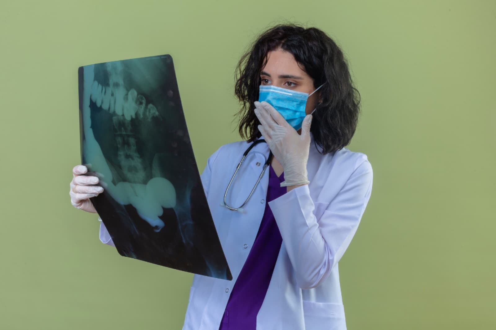
The role of imaging in healthcare has evolved dramatically, and X-rays remain one of the most widely used tools for visualising internal structures with clarity and precision. As technology has advanced, so too have safety measures, creating an environment in which diagnostic imaging is designed to be both effective and thoughtful about exposure. Many individuals naturally wonder how radiation works, how exposure is measured, and what modern systems do to keep dose levels as low as possible. Exploring these aspects offers a clearer perspective on what X-rays truly involve and how today’s imaging landscape supports informed decision-making.
What Does Radiation Exposure in Medical X-Rays Actually Mean?
Radiation exposure refers to the interaction between ionising radiation and the body during an imaging procedure. While the term can sound technical, the actual amount of radiation used in many standard X-rays is relatively low. Modern radiology equipment focuses on balancing image clarity with dose optimisation, ensuring that exposure levels align with the specific area being imaged, the type of scan selected, and the clarity required for accurate assessment.
Several factors influence exposure, including:
- The area of the body being imaged
- The density and thickness of tissues
- The number of images required
- The technique used by trained imaging staff
- The type and age of the equipment
- Whether digital or analogue systems are used
- The intended purpose of the scan
Advancements in imaging technology now allow for clearer images at significantly reduced radiation doses compared with systems from previous decades.
How Do Medical X-Ray Doses Compare with Everyday Background Radiation?
Many people are surprised to learn that radiation is not exclusive to medical imaging. Everyone is exposed to naturally occurring background radiation every day—through sunlight, soil, building materials, and even household items. Understanding this provides a helpful benchmark for placing medical imaging doses into context.
Common everyday background sources include:
- Cosmic rays from the sun
- The earth’s natural minerals
- Air travel
- Building materials
- Household ceramics and clocks
To illustrate how X-ray exposure compares with routine environmental exposure, the table below highlights approximate values:
How Do Typical Radiation Doses Compare?
| Imaging Type / Exposure Source | Typical Dose (mSv) | Notes |
| Natural background radiation (annual) | ~2–3 mSv | Everyday exposure |
| Chest X-ray | ~0.1 mSv | Low-dose imaging |
| Dental X-ray (bitewing) | ~0.005–0.01 mSv | Among the lowest exposures |
| Mammogram | ~0.4 mSv | Dose varies by system |
| CT scan (head) | ~2 mSv | Higher due to cross-sectional detail |
| CT scan (abdomen) | ~8 mSv | Varies based on equipment and scope |
These values are approximate and intended for general comparison rather than precise measurement.
Which Factors Influence Dose Levels During an X-Ray?
Not all X-rays involve the same level of radiation. Multiple variables shape the final dose, and modern imaging aims to use the lowest exposure necessary to obtain a clinically useful image.
Key factors include:
| Factor | Impact on Exposure | Description |
| Body area imaged | Moderate–High | Larger or denser regions require more exposure |
| Technology type | Low–High | Modern systems often reduce dose |
| Image quality requirements | Low–Moderate | Higher clarity can increase dose |
| Patient size/anatomy | Low–Moderate | Radiation adjusts to penetrate tissues appropriately |
| Number of images taken | Moderate–High | More images increase exposure |
| System calibration | Low–Moderate | Up-to-date calibration can reduce repeat imaging |
Newer digital imaging systems frequently integrate dose optimisation features that support high-quality imaging at reduced exposure levels.
When Are Medical X-Rays Commonly Used?
X-rays provide essential insight into structures that cannot be assessed externally. While the decision to undergo imaging depends on several considerations, the technology is used in a wide range of healthcare settings.
Common applications include:
- Reviewing chest structures
- Investigating trauma
- Assessing bone alignment
- Monitoring certain conditions
- Evaluating dental anatomy
- Supporting diagnostic planning
X-rays form part of a broader family of imaging tools, including CT scans, which use X-ray technology to produce detailed cross-sectional images.
How Does Modern Radiology Support Safe Imaging Practices?
Safety in diagnostic imaging is supported by multiple measures designed to minimise exposure while maintaining image quality. Today’s radiology teams use technology, shielding, and positioning techniques that reduce unnecessary exposure.
Common safety measures in imaging facilities include:
- Lead aprons or protective shielding
- Beam collimation to narrow exposure
- Digital sensors that require less radiation
- Routine calibration of equipment
- Protocols for reducing repeat imaging
- Dose monitoring technology
- Adjustments tailored to the patient’s anatomy
These measures are standard practice in contemporary radiology settings.
Why Does Understanding Radiation Exposure Matter?
Understanding how radiation works, the typical doses involved, and the safety measures in place helps individuals feel more informed when discussing imaging options with healthcare teams. It also highlights the importance of balancing valuable diagnostic information with thoughtful exposure management.
A broader perspective includes recognising the relative scale of exposure compared with everyday environmental radiation, while acknowledging ongoing advances in radiology technology that continue to refine safety.
How Does Border Radiology Support Safe, Modern Diagnostic Imaging?
Border Radiology stands out as a trusted provider of contemporary imaging services, offering advanced radiology technology designed to optimise both clarity and safety. The team integrates modern digital systems, dose-reduction tools, and precise imaging protocols to deliver detailed diagnostic information with an emphasis on patient care. With a combination of experienced imaging professionals, meticulous system calibration, and carefully developed imaging pathways, Border Radiology provides a reassuring environment for individuals seeking diagnostic clarity supported by thoughtful exposure management. Their commitment to ongoing technological improvement ensures access to imaging that aligns with today’s expectations for accuracy, efficiency, and safety-focused practice.
References
Radiation Health & Safety — Australian Radiation Protection and Nuclear Safety Agency (ARPANSA)
https://www.arpansa.gov.au
Radiation Exposure in Medical Imaging — ARPANSA Fact Sheet
https://www.arpansa.gov.au/understanding-radiation/radiation-sources/more-radiation-sources/medical-imaging
Radiation Dose Comparison Chart — ARPANSA
https://www.arpansa.gov.au/sites/default/files/legacy/pubs/radiationprotection/factsheets/is_medicalimaging.pdf
FAQs
- Are medical X-rays considered safe for most people?
Medical X-rays generally involve low levels of radiation. Modern equipment and imaging protocols focus on balancing image clarity with optimised exposure.
- Do CT scans involve more radiation than standard X-rays?
CT scans typically involve higher doses because they create detailed cross-sectional images. These images provide more information than a single two-dimensional X-ray.
- Is radiation from dental X-rays significant?
Dental X-rays are among the lowest-dose imaging procedures, due to the small area being imaged and modern digital sensor technology.
- How does everyday background radiation compare to imaging?
Most people are exposed to 2–3 mSv of natural background radiation annually. Many common X-rays involve far lower exposure than this yearly average.
- Can protective shielding be used during an X-ray?
Lead aprons or protective devices may be used depending on the type of imaging and the region being scanned. Their use is determined by imaging protocols.
- Why do some procedures require multiple images?
Multiple views may be required to obtain a complete picture of an anatomical region. The number of images taken is based on the information required.
This article is general information, not a substitute for medical advice. For guidance about your own situation, speak with your GP or call us on (02) 6046 9755.