

2 December 2024
Detecting Trauma and Injury with Pelvic Radiographs: A Guide to Common Findings
Learn how pelvic radiographs help detect trauma and injuries, with a guide to common findings for accurate diagnosis and treatment.
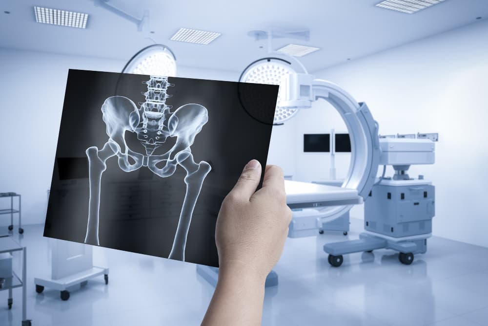
When it comes to diagnosing pelvic trauma and injuries, the role of pelvic radiographs cannot be overstated. These imaging techniques serve as critical tools in emergency radiology, allowing healthcare professionals to evaluate various types of pelvic injuries, such as fractures and dislocations. This guide aims to provide insights into the common findings observed in pelvic radiographs and the attributes that aid in accurate diagnosis.
What Are Pelvic Radiographs?
Pelvic radiographs, commonly referred to as X-rays, are essential in assessing the integrity of the pelvic ring and its surrounding structures. The most typical views include the anteroposterior (AP) view, lateral view, and inlet/outlet views. Each view serves a specific purpose, providing different perspectives on potential injuries.
Common Attributes of Pelvic Radiographs
- Radiograph Type:
- AP view
- Lateral view
- Inlet/outlet views
- Injury Types:
- Fractures
- Dislocations
- Hematomas
- Ligamentous injuries
- Diagnostic Indicators:
- Bone alignment
- Fracture lines
- Joint space narrowing
- Cortical irregularities
- Patient Positioning:
- Supine
- Prone
- Lateral decubitus
- Imaging Quality:
- Clarity
- Contrast
- Exposure settings
- Common Findings:
- Pelvic ring fractures
- Acetabular fractures
- Sacral fractures
- Risk Indicators:
- High-energy impact
- Osteoporosis
- Age-related bone density
- Radiologic Signs:
- Disrupted Shenton’s line
- Widened sacroiliac joint
- Pubic symphysis diastasis
How Can Pelvic Radiographs Help Detect Trauma?
Pelvic radiographs are instrumental in detecting a variety of pelvic injuries. The assessment process involves careful interpretation of the radiographic images to identify any abnormalities. A thorough evaluation typically follows these steps:
- Evaluate Radiographic Views: Ensure that the correct positioning for AP, inlet, and outlet views has been maintained, allowing for a symmetrical assessment of the pelvis.
- Identify Fracture Lines and Patterns: Radiologists should look for disruptions in the pelvic ring and examine the acetabulum for potential fractures.
- Assess Joint Integrity: Checking the sacroiliac joints for widening or irregularities, along with evaluating the pubic symphysis for diastasis or displacement, is crucial.
- Look for Soft Tissue Changes: The presence of soft tissue swelling or hematomas can indicate trauma, and any foreign bodies or fragments in the pelvic area should be noted.
- Consider Secondary Signs: Anomalies in Shenton’s line can suggest femoral neck fractures, while assessing the obturator foramen for asymmetry can indicate underlying injuries.
What Are the Common Pelvic Injuries Detectable on Radiographs?
Common Pelvic Injuries Include:
- Pelvic Ring Fractures:
- Lateral Compression Fractures: Often resulting from side impacts, these fractures compress the pelvis and can cause significant deformity.
- Anteroposterior Compression Fractures: Caused by frontal impacts, leading to widening of the pelvic ring.
- Vertical Shear Fractures: High-energy impacts can result in vertical displacement of the hemipelvis.
- Combined Mechanism Fractures: These involve multiple forces acting on the pelvis simultaneously.
- Acetabular Fractures:
- Anterior Wall Fracture: A fracture that occurs at the front part of the acetabulum.
- Posterior Wall Fracture: A fracture at the back part of the acetabulum.
- Anterior Column Fracture: Involves the front supporting column of the acetabulum.
- Posterior Column Fracture: Affects the back supporting column.
- Sacral Fractures:
- Transverse Sacral Fractures: These fractures traverse the sacrum horizontally.
- Vertical Sacral Fractures: Result from high-energy injuries leading to vertical disruption.
- Sacroiliac Joint Diastasis: Widening of the sacroiliac joint can indicate ligamentous injuries.
- Ligamentous Injuries:
- Sacrospinous Ligament Injuries: Can occur alongside pelvic fractures.
- Sacrotuberous Ligament Injuries: Injuries that can compromise pelvic stability.
What Are the Key Radiographic Findings in Pelvic Trauma?
Radiographic findings can significantly vary depending on the type of injury sustained. The table below outlines the different types of pelvic fractures and their key radiographic findings.
Types of Pelvic Fractures and Key Radiographic Findings
| Fracture Type | Description | Radiographic Findings |
| Lateral Compression Fractures | Side impact leading to compression of pelvis | Buckling of iliac wing, overlap of pubic bones |
| AP Compression Fractures | Frontal impact, widening of pelvic ring | Pubic symphysis diastasis, SI joint widening |
| Vertical Shear Fractures | High-energy vertical force | Asymmetry in hemipelvis, vertical displacement |
| Acetabular Fractures | Injury to hip socket | Fractures of anterior/posterior wall or column |
| Sacral Fractures | Direct impact or indirect forces | Irregularities in sacral foramina, sacroiliac gap widening |
What Are Some Common Radiographic Signs in Pelvic Trauma?
Certain radiographic signs can also assist in identifying the severity and nature of pelvic injuries. The table below summarises these signs.
Radiographic Signs in Pelvic Trauma
| Radiographic Sign | Indicator | Associated Injury |
| Disrupted Shenton’s Line | Irregular contour of femoral neck | Femoral neck or acetabular fracture |
| Widened Sacroiliac Joint | Increased sacroiliac joint space | Sacroiliac joint dislocation or ligament tear |
| Pubic Symphysis Diastasis | Separation of pubic bones | Pelvic ring instability, open book fractures |
| Asymmetrical Obturator Foramen | Uneven obturator shape | Pelvic rotation, hemipelvis shift |
| Hemipelvis Misalignment | One side of pelvis higher than other | Vertical shear or rotational injury |
What Does Research Say About Pelvic Radiography in Trauma Detection?
Studies highlight the importance of accurate imaging techniques in diagnosing pelvic injuries effectively. For instance, research published in the Australian Journal of Emergency Management outlines the critical role of timely radiographic assessments in managing trauma cases. It emphasises that understanding the radiographic features of common pelvic injuries is crucial for healthcare providers, as it influences treatment decisions and patient outcomes.
Furthermore, guidelines from the Royal Australian College of General Practitioners reinforce the significance of thorough imaging protocols and interpretation skills, ensuring healthcare professionals are equipped to handle pelvic trauma effectively.
By choosing Border Radiology Specialists, you can trust that your imaging needs will be met with the highest level of expertise and care. We are committed to delivering accurate diagnoses that play a vital role in your healthcare journey.
References
- Australian Journal of Emergency Management: https://ajem.infoservices.com.au
- Royal Australian College of General Practitioners: https://www.racgp.org.au
FAQs
- What are the common indications for pelvic radiographs in trauma cases?
Pelvic radiographs are typically indicated in cases of suspected pelvic fractures, high-energy trauma, patients with significant hip pain, or when patients exhibit signs of internal bleeding. - How do you assess the quality of a pelvic radiograph?
Assessing the quality involves evaluating the clarity of the image, ensuring appropriate contrast, and confirming that the entire pelvis is captured in the view without artifacts or blurring. - What are some signs of pelvic ring instability on radiographs?
Signs of instability include disrupted Shenton’s line, widened sacroiliac joints, and pubic symphysis diastasis, all of which can indicate severe injury. - Can pelvic radiographs detect soft tissue injuries?
While pelvic radiographs primarily assess bone structures, they can sometimes suggest soft tissue injuries through indirect signs, such as the presence of hematomas or significant swelling. - How can I prepare for a pelvic radiograph?
Patients are usually advised to wear loose-fitting clothing and inform their healthcare provider of any previous imaging or medical conditions that may affect the examination. - What role does Border Radiology Specialists play in managing pelvic trauma?
Border Radiology Specialists offer comprehensive radiographic assessments and interpretations, providing critical insights that aid in the diagnosis and management of pelvic injuries.
This article is general information, not a substitute for medical advice. For guidance about your own situation, speak with your GP or call us on (02) 6046 9755.