

2 December 2024
Advanced Imaging for Liver Disease: What Should Radiologists Know?
Discover advanced imaging techniques for liver disease and how radiologists can use them to accurately diagnose conditions like cirrhosis and liver cancer.
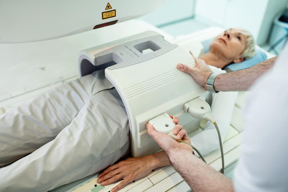
Liver disease is a significant health issue globally, affecting millions of people and leading to serious complications such as cirrhosis, hepatocellular carcinoma, and liver failure. Advanced imaging techniques have become pivotal in diagnosing, monitoring, and treating liver disease, providing radiologists with critical insights into liver morphology and functionality without invasive procedures. This article explores essential aspects of advanced imaging in liver disease, diving into the various imaging modalities, key parameters for assessment, and the clinical applications of each technique.
Why is Advanced Imaging Essential for Liver Disease?
Advanced imaging offers non-invasive, accurate means to assess liver health, fibrosis progression, and the presence of tumours or fatty infiltration. With modalities such as MRI, CT, and ultrasound (including elastography), radiologists can effectively diagnose conditions such as cirrhosis and hepatocellular carcinoma. Imaging also helps evaluate vascular changes due to portal hypertension and quantifies liver fat in conditions like non-alcoholic fatty liver disease (NAFLD). By providing detailed images and quantifiable data, these techniques have reduced the need for liver biopsies, making the assessment process safer and less invasive for patients.
Which Imaging Modalities are Best for Liver Disease?
Each imaging modality offers specific benefits and limitations, making them suitable for particular aspects of liver disease. Here’s a breakdown:
| Imaging Modality | Advantages | Limitations | Common Uses in Liver Disease |
| Ultrasound | Non-invasive, widely available | Operator-dependent, limited in obese patients | Initial screening, focal lesion assessment |
| CT | High spatial resolution, fast | Ionizing radiation, not ideal for soft tissue | Tumour staging, vascular assessment, trauma evaluation |
| MRI | Excellent soft tissue contrast | Expensive, long acquisition time | Fibrosis staging, lesion characterisation, steatosis quantification |
| Elastography | Non-invasive fibrosis assessment | Limited accessibility | Fibrosis staging, portal hypertension assessment |
| Nuclear Medicine | Functional information | Low spatial resolution | Tumour characterisation, staging |
What Parameters are Used to Assess Liver Disease?
Advanced imaging in liver disease incorporates several key parameters that assist in evaluating the extent and nature of the disease. The primary parameters are:
- Liver Fibrosis: Grading and staging of fibrosis using elastography (e.g., MR Elastography or ultrasound-based techniques).
- Steatosis (Fatty Liver): Quantification of liver fat content, commonly through MRI Proton Density Fat Fraction (PDFF) measurements.
- Inflammation and Necrosis: MRI sequences identify inflammation levels, crucial for assessing chronic hepatitis or alcoholic liver disease.
- Tumour Identification and Characterisation: Differentiating between benign and malignant liver lesions, which is especially critical in hepatocellular carcinoma (HCC) cases.
- Portal Hypertension: Detection of vascular changes via Doppler ultrasound or CT angiography to assess elevated portal pressures, a common complication in advanced liver disease.
Which Imaging Techniques Are Used for Different Types of Liver Disease?
| Liver Disease Type | Preferred Imaging Modality | Reason for Preference |
| Hepatocellular Carcinoma | MRI, CT | High sensitivity and specificity for lesion detection |
| Liver Fibrosis | Elastography (MRE, Ultrasound) | Non-invasive, allows grading of fibrosis without biopsy |
| Fatty Liver Disease | MRI PDFF | Accurate quantification of fat content |
| Cirrhosis | Ultrasound, MRI | Early detection of nodularity, vascular changes |
| Vascular Disorders | Doppler Ultrasound, CT, MRI | Enables assessment of blood flow and vascular structures |
How Does MRI Aid in the Assessment of Liver Disease?
MRI is considered the gold standard in liver imaging due to its superior soft tissue contrast and versatility. Gadoxetic acid-enhanced MRI is particularly valuable for assessing liver function, as it provides enhanced visibility of liver parenchyma and lesions. MRI sequences such as T1 and T2-weighted imaging, diffusion-weighted imaging, and MR elastography allow for comprehensive analysis:
- Fibrosis and Cirrhosis: MR elastography assesses liver stiffness, which correlates with fibrosis severity. This non-invasive technique offers a reliable alternative to liver biopsies for staging liver fibrosis.
- Fatty Liver Disease: Proton Density Fat Fraction (PDFF) MRI quantifies fat levels within the liver, enabling early detection and monitoring of NAFLD and NASH (non-alcoholic steatohepatitis).
- Tumour Identification: With multiphase imaging, MRI allows radiologists to distinguish between malignant and benign lesions, providing critical insights into hepatocellular carcinoma (HCC) and metastasis.
What Role Does CT Play in Liver Disease Imaging?
CT is widely used for liver disease imaging, particularly in emergency settings and for assessing structural and vascular changes. Multiphase CT is especially helpful for detecting hepatocellular carcinoma by capturing arterial, venous, and delayed phases. Dual-energy CT provides additional benefits in differentiating tissue types and assessing fatty infiltration. Although CT involves ionising radiation, its high resolution and speed make it an excellent choice for:
- Tumour Staging: CT is ideal for staging liver cancer and identifying metastasis in surrounding tissues.
- Vascular Assessment: Contrast-enhanced CT is commonly used to assess portal hypertension and hepatic vein thrombosis.
- Trauma Evaluation: For patients with traumatic liver injury, CT helps determine the extent of damage and plan treatment.
How Do Elastography Techniques Contribute to Liver Disease Assessment?
Elastography has emerged as a powerful, non-invasive tool for liver stiffness measurement, which correlates with fibrosis and portal hypertension. There are several types of elastography:
- Transient Elastography (TE): Using FibroScan, this technique measures liver stiffness and is widely available in clinical practice.
- Shear Wave Elastography (SWE): Performed with ultrasound, SWE provides quantifiable liver stiffness and has proven useful for liver fibrosis assessment.
- Magnetic Resonance Elastography (MRE): MRE uses MRI technology to assess liver stiffness, offering a high level of accuracy and consistency across patients.
Each of these techniques reduces the need for liver biopsy in many cases, making liver disease diagnosis safer and more accessible.
How is Nuclear Medicine Applied to Liver Disease Imaging?
In liver disease, nuclear medicine, specifically PET/CT, is used less frequently but can be valuable for tumour characterisation and staging. PET/CT combines metabolic and anatomical data, making it useful for evaluating metastatic liver disease or residual cancer after treatment. It’s especially relevant for patients where other imaging modalities have not provided definitive results.
What Clinical Uses Does Advanced Imaging Offer in Liver Disease?
Advanced imaging in liver disease has a variety of clinical applications:
- Diagnosis of Fibrosis and Cirrhosis: Non-invasive staging of liver fibrosis through elastography and MRI.
- Staging of Hepatocellular Carcinoma (HCC): CT and MRI detect and characterise liver tumours, including HCC and metastases.
- Evaluation of Portal Hypertension: Doppler ultrasound and contrast-enhanced CT assess elevated portal pressures.
- Quantification of Liver Fat (Steatosis): MRI-PDFF enables quantification of fat content in NAFLD cases.
- Pre- and Post-Treatment Assessment: Imaging helps monitor liver disease progression and response to treatment over time.
At Border Radiology Specialists, we are dedicated to providing exceptional liver imaging services, including the latest advancements in elastography, CT, MRI, and PET/CT, ensuring optimal care and insights for liver disease diagnosis and management.
References
- Australian Liver Association. (2023). Liver Disease and Imaging Guidelines. Australian Liver Association.
- Hepatology Research Group. (2022). Advanced Imaging Techniques in Liver Disease. University of Queensland Press.
- World Journal of Gastroenterology. (2021). “MRI and Elastography for Non-Invasive Liver Disease Assessment”. World Journal of Gastroenterology.
Frequently Asked Questions (FAQs)
What is the best imaging modality for diagnosing liver cancer?
MRI, particularly with Gadoxetic acid enhancement, is highly effective for diagnosing liver cancer due to its superior soft tissue contrast and sensitivity to lesion characterisation.
How does elastography work in assessing liver fibrosis?
Elastography measures liver stiffness, which correlates with fibrosis severity. Techniques like MRE and transient elastography (FibroScan) offer non-invasive alternatives to biopsies.
Why is MRI preferred over CT for fatty liver disease?
MRI’s Proton Density Fat Fraction (PDFF) sequence can accurately quantify liver fat, making it ideal for early diagnosis and monitoring of fatty liver disease.
Can advanced imaging detect early stages of cirrhosis?
Yes, advanced imaging such as MRI and ultrasound elastography can detect early signs of cirrhosis, including changes in liver stiffness and nodularity.
Is nuclear medicine commonly used for liver imaging?
Nuclear medicine is less common but valuable for tumour staging and metabolic assessment when other modalities are inconclusive, particularly in metastatic liver disease.
How does Doppler ultrasound help in liver disease?
Doppler ultrasound is essential in assessing blood flow in the liver and detecting vascular complications, such as portal hypertension, often associated with liver disease.
This article is general information, not a substitute for medical advice. For guidance about your own situation, speak with your GP or call us on (02) 6046 9755.